|

Kyle Perry
|
Previous studies have shown that
the mammalian cell cytosol is generally non-permissive for the growth
of bacteria, which suggests that intracellular bacterial pathogens such
as L. monocytogenes have
evolved specific mechanisms to allow for growth in this environment.
However, relatively little is known about the mechanisms facilitating
intracellular survival, growth, and spread of bacterial pathogens. To
identify bacterial factors that allow intracellular infection by L. monocytogenes, I am employing
two fluorescence-activated cell sorting (FACS)-based genetic screens.
To identify bacterial factors
specifically involved in cell-to-cell spread, murine bone
marrow-derived
macrophages (BMM) are infected with a transposon library of
GFP-expressing L. monocytogenes.
After several hours of infection, host cells are processed via FACS to
enrich for BMM with high fluorescence, which contain transposon mutants
with cell-to-cell spread defects. To identify bacterial factors
involved in intracellular replication, BMM are infected with a
transposon library of ActA-deficient
L.
monocytogenes expressing GFP. ActA-deficient bacteria are defective for
intracellular actin-based motility and cell-to-cell spread. Infected
host cells are similarly processed via FACS to enrich for BMM with low
fluorescence, which contain transposon mutants with intracellular
replication defects. Cell-to-cell spread and intracellular replication
defects of mutants identified in both screens are being characterized
by genetic analyses and intracellular infection models.
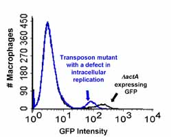
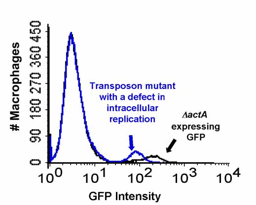
View Larger Image
Figure. Flow cytometry analysis of
infected bone marrow-derived macrophages (BMM). Exponential phase
bacterial cultures were used to infect BMM at an MOI of 1. After
6 hr, the infected macrophages were analyzed by flow cytometry. The
black line represents the fluorescence intensity of macrophages
infected with an ActA-deficient strain of L. monocytogenes expressing GFP.
The blue line represents the fluorescence intensity of macrophages
infected with a transposon mutant of the GFP-expressing ActA-deficient
strain that results in a defect in intracellular replication.
|
